Baseline ECG changes in life-threatening arrhytmia

When assesing patients with collapse with unidentified cause, its possible underlying arrhytmia is present. The following ECG changes can suggest this

  • 1. Short PR interval and delta-wave: WPW
  • 2. Epsilon wave in arrhytmogenic (right) cardiomyopathy
  • 3. Long QT syndrome
  • 4. Brugada sign (type 1 or type 2)


This list is ongoing and not complete yet

How to visualize cardiology patients

For every cardiology patient visualise the status of the following stuff:

  1. Electrical Rhythm and conduction
  2. Coronary arteries
  3. Heart muscle function
  4. Valve function
  5. Important accesories: pericard, vasculator (aorta), systemic diseases affecting the heart (DM2), elektrolytes, pulmonary function

1. Electrical Rhythm and conduction

Tachyarrhytmias, bradyarrhytmias, heart blocks, pacemakers

2. Coronary arteries

Coronary artery disease, risk factors, Stress-test/angiograms/scan?, PCI or CABG history?

3. Heart muscle function

Cardiomyopathies,

4. Valve function

Regurgitation, stenosis

5. Accesories

pericard, vasculator (aorta), systemic diseases affecting the heart (DM2), elektrolytes, pulmonary function

Chest pain: Boerhaave syndrome

Case: A 50 year old male with alcohol abuse present in the Emergency department with mild chest pain. He has been nautious and vomiting the past couple of days. Also there is diffuse abdominal tenderness. He gets admitted to control pain and CT-scan next days but detiorates suddenly. He dies. Obduction shows a ruptured oesophagus.

Boerhaave syndrome is rupture of esophagus and rapidly fatal.

History: a dutch grand admiral was retching and tore his oesophagus apart. He died after eating a duck.

Pathofysiolygy: rupture of esophagus due to increase intraluminal pressure usual due to vomiting or retching. Alcohol intake is related. Higher risk if gastro-intestinal disease is present.

Other causes: straining, childbirth, heavy lifting, seizures, fits of coughing or hiccup

Clinical symptoms: chest pain, nautious, vomiting (bloody), dysphagia, dyspnea,

Anderson Triad: Subcutaneous emphysema, rapid respirations, and abdominal rigidity

Macler triad: vomiting, pain, subcutaneous emphysema

Physical examination: Crepitus in the neck, abdominal rigidity, abdominial pain. tachycardia, tachypnoe
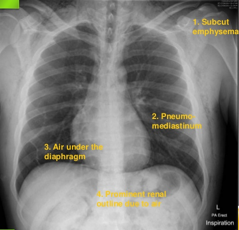
Chest X-ray signs:

  • Subcuteneous emphysema
  • Pneumomediastinum
  • Free air under the diaphragm
  • Pleural fluid

boerhaave

If still suspected make CT-thorax/abdomen (contrast in esophagus?).

Treatment:

* Surgery as soon as possible

Aggresive resuscitation and broadspectrum antibiotics.

The cause of death is usually polymicrobial septic shock and mediastinitis.

Differential diagnosis of chest pain


Serious: STE-ACS, dissection, thoracal aneurysm, massive pulmonary embolism, pericardtamponade, tension pneumothorax, esophagel rupture


Moderate: NSTE-ACS, pulmonary embolism, pericarditis, pleuritis, pneuomnia, unstable angina pectoris, aortic stenosis, pneumothorax,


Mild: tendomyogenous, ribfracture, reflux, panic, stable angina pectoris, hyperventilation, costochondritis,


Gastro-intestinal: Pancreatitis, stomach ulcus, gallstones

Diagnostic consideration: D-dimer, abdominal enzymes, X-thorax

  • Females and diabetics have aspecific chest pains when enduring myocardial ischemia